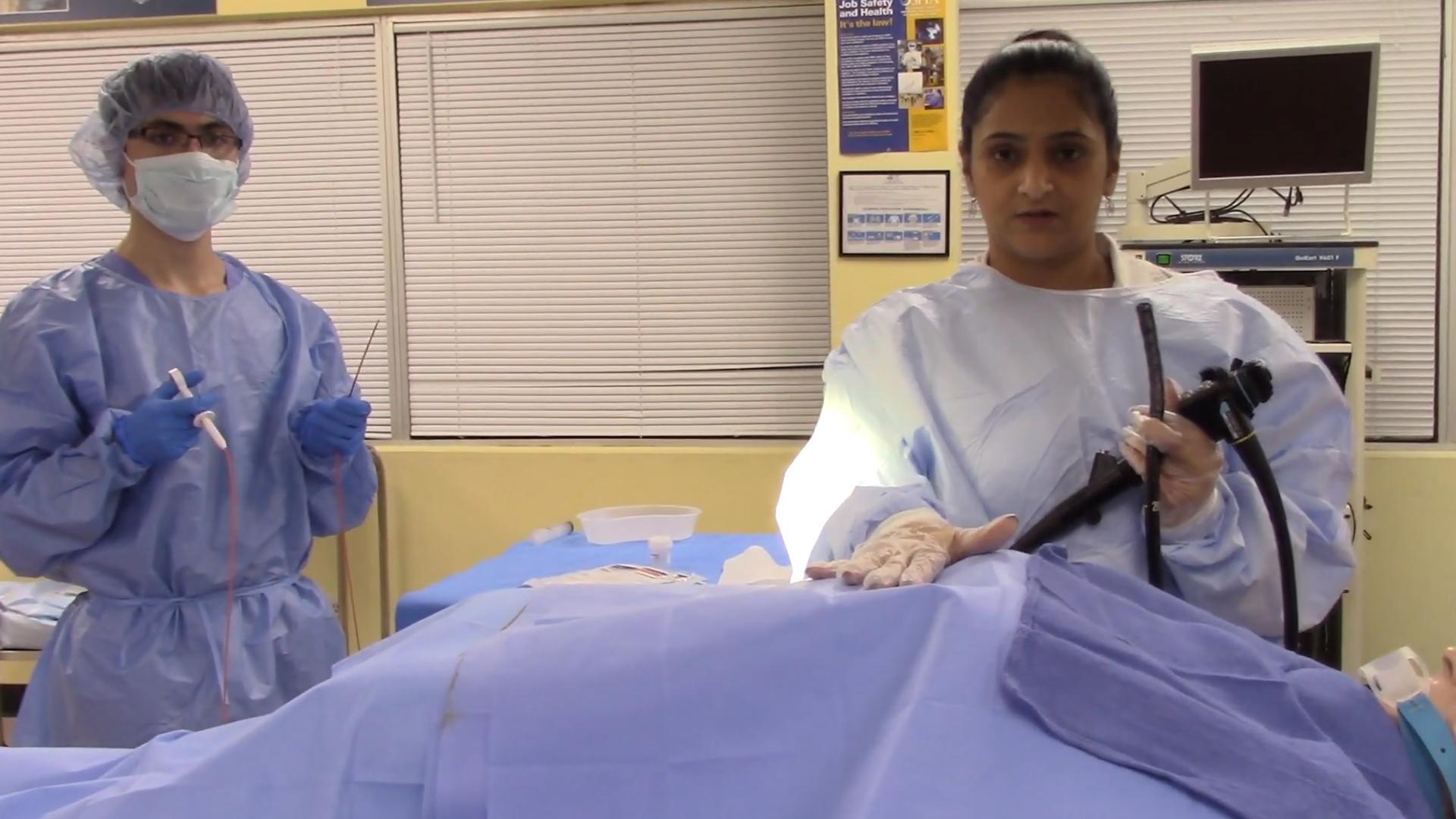
Gastroscopy is a medical diagnostic procedure. The inside of the oesophagus, stomach, and first section of the intestinal tract can be examined using a narrow tube (an endoscope). During the study, the tube includes a light and a camera on the end that delivers images to a monitor. As a result, a gastroscopy in gastroscopy clinic may be performed to identify disorders involving the oesophagus, stomach, and duodenum.
- Gastroscopy is done with an endoscope, which is a thin flexible tube with a tiny camera and, in some situations, light at the end. This camera is designed to capture photos of the oesophagus, stomach, and duodenum.
- The tube is introduced via the mouth and directed down the neck into the oesophagus and then stomach for around 15 minutes. Although the operation is not uncomfortable, a numbing spray could be used to relax the neck somewhat before inserting the tube.
- A gastroscopy in gastroscopy clinic would indeed be performed to examine for problems in the duodenum and stomach. Tissue samples can be obtained for biopsies, polyps can be removed, and the presence of certain bacteria, such as the bacterium H pylori, which causes many peptic ulcers, can be determined. Bleeding ulcers can potentially be cauterised during the gastroscopy operation.

- Preparing for a gastroscopy: Certain drugs may be discontinued in the weeks coming up to a gastroscopy. The stomach has to be empty; no solid food from of the previous night may be consumed, and only water may be consumed. It is also critical not to smoke well before exam.
- The scan is not normally painful. It does, however, produce nausea when the tube is put into the throat, although a freezing spray can be used to make it a little less painful. A gastroscopy may always be required if any abnormalities are discovered since samples or biopsies of the intestines or polyps cannot be extracted during a barium test.


